
Molecular and Cellular Scene for a Journal Cover
Process

Sketches
We first mocked up sketches to illustrate the main components and layout, taking into consideration the placement of the journal cover text.

Visualization of the Protein Structures
The structures of the LDL-Receptor protein and viral surface glycoprotein were accurately visualized utilizing structural data from crystallized protein domains.
Model of LDL-R (right panel)

Visualization of Protein M Mutant
The featured protein was represented by a predicted model for the M51R mutant matrix (M) protein.The critical methionine to arginine substitution at residue 51 was highlighted.
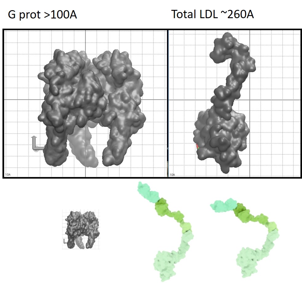
Molecular Size Comparison
To maintain accuracy the atomic sizes of the molecules were determined and proteins were scaled accordingly.

Color Palette Selection
We shared several options for the final color palette to ensure we could arrive at the desired look and feel. Can you tell that these options were inspired by the ideas of hero versus villain, and space travel?

Initial Rendering of the Protein Structures

Final Journal Cover Image
Our final image features realistic 3D representations of the Vesicular Stomatitis Virus M51R mutant protein and a colon carcinoma cell. The accurate visualization of the protein structures were achieved through the use of structural data from X-ray crystallography, supplemented by comparative homology and ab initio protein structure prediction methods. The placement of light and use of color communicate the potential positive therapeutic impact of the M51R mutant VSV for colon carcinoma treatment.